


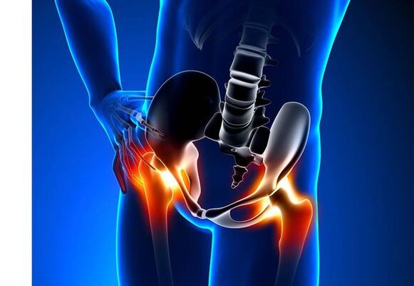
The hip joint is arthrosis (coxarthosis)- This is a chronic degenerative joint disease that leads to deformation of bone tissue.With coksartrosis, all components of the joint are involved in the pathological process: joint cartilage, bone structures next to the cartilage, synovial shell, ligaments, capsules and adjacent muscles.In the case of illness, the articular cartilage is destroyed, the micro reduction of the bones and osteophytes (bone growth) appears, and inflammation of the muscle ligaging of the hip.
One in five people in the world complains of joint problems with joints.It can be both pain, or restricting movement in the joints and can be a combination of these symptoms.Every second outpatient vision applies to patients with bone-to-muscular disorders, while 66 % of cases are people under 65.According to the latest epidemiological research, the prevalence of arthrosis of the knee and hip joints between the adult population is 13 %.
Risk factors for the development of coxarthrosis:
- Genetic predisposition.The general cause of cocsartrosis of the hip joints is described in II.Type or acquired mutation of type of type prollagen type.
- Elderly.The likely reason for the prevalence of arthrosis in the elderly is the harmful effect on the joint cartilage and the ability to restore.
- Floor.Women suffer more often in osteoarthritis than men.The reason for this is that the effect of estrogen female sex hormones on bone mineral metabolism.However, the influence of the floor is unclear - according to some authors, with the damage to the other joints, there is no difference in sexual sexual sexual: in men, the hip arthrosis is found as often as in women.
- Unnecessary body weight.The connection has been proven between body weight and the occurrence of arthrosis.Excess adhesive tissue increases the harmful load on the cartilage.In addition, adipose tissue produces pro -inflammatory enzymes that damage the cartilage tissue.
- Frequent development of bones and joints.In accordance with the tests, 80 % of coxarthrosis, which occurs for no obvious reason, is related to previously non -diagnosed errors in the development of hip joint and subluxation.
- Hard physical work.Excessive burden on hip joints with certain types of physical birth can lead to joint damage and joint development.There are endangered agricultural workers, spades and people with similar workers.
- Injuries.The risk of developing coxarthrosis increases after injury to the hip joint.In fact, both injured joints, both of which can participate in the process.
- Professional sports.Professional sports can provoke the occurrence of coxarthrosis both due to excessive joints and injuries.Potentially dangerous sports include heavy athletics, athletics jump, parachute sports.
- Bones and joint diseases- rheumatoid arthritis, psoriatic arthritis, joint infections, avascular necrosis, gouty arthritis, etc.
- Endocrine pathologies- hypothyroidism, hypoparathyroidism, acromegaly (anterior part of the pituitary gland), diabetes, obesity.
If you notice similar symptoms, consult a doctor.Don't meditate yourself - dangerous to your health!
The joints of the hip joints
The main symptoms of coxarthrosis are: pain, mobility restrictions and crunching in the joints, their deformation, functional shortening of the lower limb and periodic swelling of the joints.
Pain of different intensity.The pain in the joint is initially insignificant and occurs for a short period of time.They appear or increase during walking or with other physical efforts such as squatting, tendency and weight lifting.With the development of the disease, the pain increases and even a long rest does not bring relief.In addition, pain is accompanied by prolonged mobility of the joint and fixing in one position.
Patients complain of so -called "beginner" pains in the hip joints after sleeping, driving in a car and other prolonged mobility.The "beginner" pain for coxarthrosis lasts up to 30 minutes.The pain increases during hypothermia or stressful position.They can be localized in the area of the buttocks or groin, on the front or side of the thigh.With the spread of pain, the nerves of the lumbar plexus may spread to the center of the body or from the knee.Pain sometimes refers to the lumbosacral spine and the tailbone.
Restriction of joint mobility.The movement of the hip joint with coksartrosis is limited due to pain.At the same time, the rotation (turns inside and outside) and the lower limb (movement in the center of the body) are often disturbed but limited (movement from the center axis of the body), as well as bending and extension.The inability to perform passive movements in the joint is caused by compensating pools due to the pronounced pain syndrome.The patient's walk changes, the buttocks retreat, and the body progresses as it transmits the weight to the injured side.Bilateral damage to patients with coksartrosis develops "duck walking".
Coxarthrosis occurs regularlyswelling in the jointwhich can be invisible because of the muscle and the fat layer.The disease is also typicalCrystals in the joints while moving, gradual deformation and functional shortening of the lower limb.
The disease often affects a joint and then the process applies to others.But sometimes arthrosis affects several joints at a time and polyosostoarthritis occurs.Polyosteoarthrosis is characterized by bones, joints and endocrine disorders with the elderly or inherited and concomitant diseases.
The pathogenesis of arthrosis of the hip joints
Mechanical harmful (injuries and microtraumas due to increased physical efforts of the joint), as well as genetic, hormonal and metabolism, play an important role in the pathogenesis of hip joints.It is often not possible to find out which factor has affected the disease of the disease, but the disease often develops after mechanical injuries after tissue damage.
Damage to the tissue stimulates the division of cartilage cells (chondrocytes), while the production of pro -inflammatory cytokines is increasing, which are usually only small amounts of cartilage.Cytokines trigger the inflammatory procedure, for example, IL-1 anti-inflammatory cytokine can be distinguished by enzymes that destroy the cartilage of the joint.In addition, cytokines have the production of Tsog-2 enzyme and other substances that have toxic effects on the cartilage.
The Synovites also play a major role in the inflammatory diseases of the synovial shell of the synovial shells of joints or leagues, with the accumulation of fluid in the cavity.
Decrease in the elasticity and strength of joint cartilage leads to a reduction in mechanical stress resistance to metabolic disorders.In the case of coksartrosis, all components of the joints are involved in the pathological process, including a subchondral bone.Because the large joints of the lower limbs make up the large joints of the body, they experience significant mechanical tensions that cause microvices to occur on the subchondital plate and in the cartilage.As a result of microusomas, the subchondral bone is compacted, leading to regional growth of bone tissue - osteophytes.And this, in turn, stimulates further degradation of joint cartilage.
In some cases, arthrosis of the hip joint is inherited.Hereditary arthrosis is said to have a polygenic heritage - due to the effects of many genes, all of which are poorly affected.The cause of some diseases is the mutation of genes encoding the macromolecules of the articular cartilage, which causes the rupture.The genes responsible for sharing of the chondrocites may also suffer.In addition, metabolic disorders are inherited, such as pyrophosphate artropathy -a disease in which calcium -pyrophosphate crystals accumulate in the articular cartilage and synovial fluid.
Classification and stages of the development of arthrosis of the hip joints
Depending on the causes of the disease, coxarthrosis is divided into two main forms: primary (idiopathic) and secondary (because of or because of other diseases).
Primary Coksartrosis:
- Localized (affected only by hip joints):
- one -sided;
- bilateral.
- With a general (polyosteoarthrosis) lesion of at least three joint groups (such as hips, knees and small brushes or legs).
Secondary Arthrosis:
- Post -traumatic:
- Acute - acute injury;
- Chronic - Classes of some sports or as a result of professional activity.
- Metabolic Diseases (Oconis, Hemochromatosis, Wilson -Kór, Gauccher Disease).
- Congenital pathologies and developmental defects (congenital dysplasia of the hip joint, pertinent disease, epiphyse slide of the femur, hypermobility syndrome, shortening of the lower limb, skoliosis, bone disdlasia).
- Endocrine pathologies (acromegaly, hypothyroidism, diabetes -breast, hyperparatireoidism, obesity).
- Calcium salts (pyrophosphate artropathy, whitewashing tendonitis).
- Diseases of bones and joints (rheumatoid arthritis, psoriatic arthritis, pediatric disease, avascular necrosis, infections).
According to clinical manifestations, the following forms of coxarthrosis are distinguished:
- Small symptom.
- Manifested with clear, clear clinical symptoms:
- rapidly progressive, in which symptoms develop in the first four years from the onset of the disease;
- Slowly progressive - clinically significant symptoms occur after five years of the disease.
According to the X -Gay image, two joint joints of the hip joints can be identified:
- Hypertrophic - with signs of increased reparative response (injuries are replaced by new tissue, such as osteophytes);
- Atrophic (decrease in tissue volume).
The stages of the disease can be determined from a radiological and clinical point of view.The classification of Kellgren and Lawrence (1957) is most often used to determine the radiological stage of the hip joint arthrosis.
Stages of arthrosis in radiological classification
| Stage | Signs |
|---|---|
| 0 | X -try images do not have signs of arthrosis |
| 1 | The joint gap does not change, the only regional osteophytes are displayed |
| 2 | The joint gap does not change, significant regional osteophytes are displayed |
| 3 | The height of the joint gap decreases moderately, with significant regional osteophytes |
| 4 | The height of the joint gap is significantly decreased, with significant regional osteophytes and subchondral osteosclerosis (compression of bone tissue under the lower surface of the cartilage, with the structure of the cartilage) |
Classification (1961) is used to determine the clinical stage of the disease, which uses both clinical symptoms and display criteria.
The joint clinical stages
| Stage | Signs |
|---|---|
| 0 | The joint gap is clearly and unevenly narrowed, the edges of the joint cracks are slightly pointed (initial osteophytes), a slight restriction of movements |
| 1 | The joint gap is significantly narrowed (50-60 %), significant osteophytes, subchondral osteocosclerosis and cystic enlightenment in the bone epiphysees;The clinic is dominated |
| 2 | deformation, stiffness of the joint;The joint gap is narrowed or absent by more than 60-70 % of norms |
Joint complications of hip joints
In coxarthrosis, all complications are related to the abnormal changes of the joints.
The course of coksartrosis is complicated with local inflammatory processes:
- Bursite - inflammation of synovial bags in the joints;
- Tendovaginitis - inflammation of the inner shell of muscle tendons;
- The nervetunnel syndrome syndrome syndrome syndrome syndrome syndrome syndrome syndrome syndrome syndrome syndrome-syIndrome syndrome syndrome syndrome syndrome syndrome syndrome syndrome syndrome syndrome syndrome syndrome.
With the progression of coxarthrosis and the transition to clinical stages II and III, pain restricts the joint mobility and, over time, with the full agility of joint ankylosis (fibrous, bone or cartilage).
Can lead to significant joint deformationFracture of bones or aseptic necrosis.In the case of coksartrosis, the necrosis of the femur is the most fearful complication.
Can occur in case of pronounced coksartrosisSubluxation and displacement of the jointand penetration of the femur head into the pelvis cavity.Dislocation and subluxation of the hip joint leads to pain (first acute, then boring and sore), enhancing walking and other physical efforts, as well as shortening of the joint, lame, and sometimes the affected limb.
Despite the fact that in modern clinical practice, there is no systematic manifestations of joint systematic manifestations, more attention is paid to related diseases.These are pathological conditions that exist or arise in the background of the current disease.In connection with the inflammatory reactions that arise during arthrosisCardiovascular disease- a reduction in physical activity due to restraint of pain and joint mobilityObesity, depression and deterioration in quality of life-No -steroid anti -inflammatory drugs for prolonged use,The upper gastrointestinal sections are affected,And tooIncreases the risk of cardiovascular pathologies and kidney diseaseOr
Diagnosis of joint joints of the hip joints
The diagnosis of "coksartrosis" is performed on the basis of clinical manifestations and radiological examination.There are no typical laboratory signs for joint diagnosis.
Between clinical manifestationsThe main pain and character are the main diagnosis of arthrosis of the hip joint.The pain in the arthrosis of the hip joint occurs and gradually increases over several years (sometimes for several months in progressive form).The pain occurs or increases in a physical effort or constant position.If the patient feels pain alone, the inflammation (synovitis) is connected.The statement is taken into account for 30 minutes and with longer agility.
The restriction of joint mobility is gradually increasing, which applies to both active and passive movements.As the disease develops, the joints are deformed and a functional abbreviation for the limb length can occur.
On a physical examinationLimit joint mobility, their deformation, shortening of the limbs, pain in joint palpation and high spinning of the femur, muscle atrophy.
Laboratory methodsIt is not necessary to diagnose arthrosis of the hip joints.However, they can be used for the differential diagnosis of coxarthrosis with arthritis (rheumatoid and chronic), since there is no inflammatory change in the general blood test and rheumatoid factor in arthrosis and the uric acid levels do not increase.In addition, laboratory tests are contraindicated in drug treatment methods.
Instrumental methodsTo diagnose arthrosis of the hip joints:
- Radiography- This is the main method of diagnosing arthrosis of the hip joints.X -rays determine the changes typical of cocsartrosis: the narrowing of the joints, osteophate, erosion and cartilage, alchondral cysts and osteosclerosis.The X -Ray study is a classic method for diagnosing coxarthrosis and the classification of coxarthrosis.At present, however, other methods of displaying joints are increasingly being used, such as ultrasound and magnetic resonance imaging.
- Ultrasound examination (ultrasound) -The advantage of ultrasound is that the body is in the absence of radial load.
- Magnetic resonance tomography (MRI)- In comparison with other methods, this allows for a clearer display of joint damage.
- ArthroscopyOrAllows you to identify the damage to the articular cartilage: from the condomic zones (softening of the articular cartilage) to deep cracks less than 10 mm in diameter that penetrate the subchondral bone and the formation of deep ulcers.Superficial and medium cracks and surface erosion can also be displayed.
Identification of coksartrosis is usually not special difficulties, but when assessing a particular clinical situation, it should be remembered the possible secondary origin of the arthrosis of the hip joints (such as complications of other diseases such as endocrine disorders).
Treatment of arthrosis of hip joints
Treatment of arthrosis of the hip joints can be conservative (medication and not united) or functional.Conservative treatment is used in 1-2 stages of the disease and 3 stages of surgery.Surgery treatment can be recommended in 2 stages with persistent pain and lack of reaction to conservative therapy.
The purpose of conservative therapy is:
- Improve quality of life - reduce pain and increase joint mobility;
- Stop or slow down the disease.
Nem -drog treatment methods include:
- unloading of the hip joint (decrease in body weight, creating additional support and transferring part of the body weight to reed or crutches);
- physiotherapy physical education;
- Physiotherapy methods.
Treatment of coxarthrosis begins with non -drog methods, and physiotherapy exercises play an important role.In case of severe pain, the patient should use support.Disease and endoprostany contraindications should be used for life.
Cuxartrosis treatmentIt includes drugs that reduce the symptoms of the disease.These are medicines for painkillers and groups of non -steroid anti -inflammatory drugs (NSAIDs).NSAIDs are not divided into elections and selective.
Analgesic arthritis and NSAIDs are used for a short time to relieve pain and inflammation.Currently, one of the non -steroid anti -inflammatory drugs is not a proven advantage, so the choice of a particular drug depends on the side effects and the specific clinical situation caused by the cause.
It should be remembered that NSAIDs have many side effects.If you take them, it will affect the membrane of the stomach and duodenum, which results in ulcers and bleeding.Many NSAIDs have toxic effects on the liver and kidney.In addition, NSAIDs disrupt the platelet aggregation and, as a result, the patient is disrupted by thrombosis and is prone to bleeding.Long -term NSAIDs suppress the processes of hematopoiesis and can cause aplastic anemia and agranulocytosis.Receiving selective NSAIDs causes significantly less complications.
Locally used ointment and gels cause less side effects than oral products.Drugs with warming and reduction pain are used to treat arthrosis.They can contain turpentine, menthol, nicotinic acid esters, salicylates, uterine venues.In addition, NSAIDs have a good effect.
In the absence of an analgesic and NSAID, or if it is impossible to choose the optimal dose of the drug, the central action of the analgesics is short -term.
In the case of inflammation, intraarticular administration of corticosteroids is used.Corticosteroids are used up to 2-3 times a year as it can lead to more frequent use of cartilage degeneration.
Slow -acting drugs weaken the symptoms of the disease, the compounds of chondroprotectors, inadequate avocado or soy, hyaluronic acid.These drugs are included in the European Anti -Medematic League recommendations to treat arthrosis of the hip joints.Preparations reduce pain and improve joint mobility.
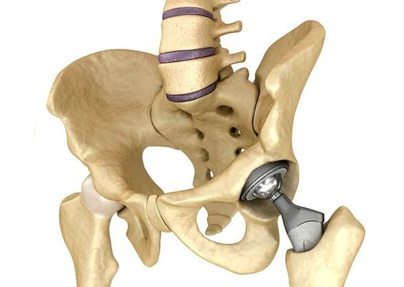
Endoprostany of the hip jointsSerious III.It is used in stages when the pain syndrome cannot be removed and the joint mobility is significantly limited.The prosthetics of the hip joint leads to a decrease in pain syndrome, to improve the functional condition of the joint and the quality of the patient's life.The effect remains for 10-15 years, then a second operation may be required.During surgery, the hip joint is replaced by artificial imitation of ceramic, metal (most often titanium prostheses) or polymer.
Forecast.Prevention
The prognosis of the arthrosis of the hip joints relative to the patient's life is favorable, but the disease often leads to disability.According to the World Health Organization, 80 % of elderly patients with coxarthrosis violate mobility and 25 % cannot do everyday matters.In this respect, primary prevention of arthrosis of the hip joints is important.
Prevention measures:
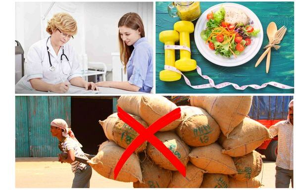
- Reduce body weight- Nutrition should be set to reduce the weight and load of the joint.In addition, the reduction of adipose tissue reduces the amount of inflammatory mediators released.
- Avoid heavy physical work and sport overload.Physical overload is often the cause of arthrosis of the hip joints, while moderate physical activity, on the contrary, improves the condition of the joint cartilage, retains its normal mobility and reduces the burden on other joints.
- Correct the underlying disease.If the patient is detected in diseases that can lead to secondary cocsartrosis (endocrine, rheumatism and others), the underlying disease is required.Normalization of the hormonal background and achieving long -term remission of rheumatic diseases are all primary prevention and allows for slowing down its development.
- Lead a healthy lifestyle.A balanced diet, with sufficient plant and animal protein, multiple unsaturated fatty acids and reduced carbohydrates, and moderate physical activity, avoid coxarthrosis, even in the presence of risk factors.
Currently, preventing the diseases of the hip in neonatology and pediatrics are mandatory.Over time, the hip joint adjusted congenital dysplasia significantly reduces the risk of adult coxarthrosis.